Author: Petr Šícho | Supervisor: Victoria Fay | Advisor: Prof. Christophe Moser (EPFL, LAPD)
Project Overview & Objective
- Motivation: Head and Neck Cancer (HNC) affects ~1M people/year. Real-time tumor margin mapping is critical to ensure complete surgical resection and prevent recurrences.
- Approach: Developing a machine learning segmentation algorithm (U-Net variants) leveraging Fluorescence Lifetime Imaging Microscopy (FLIM) to classify tumor cells via endogenous metabolic signatures (Warburg effect).
Methodology & Data Acquisition
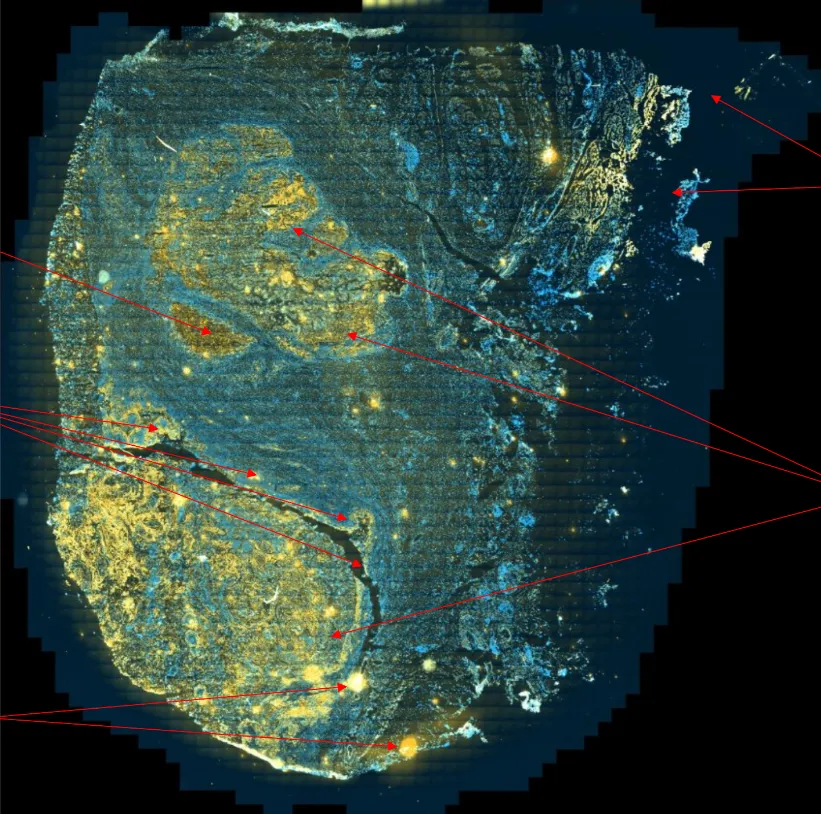
- Data Gathering: Captured FLIM and intensity data across two wavebands (NADH/FAD cofactors) from human lymph node sections using a Leica SP8 microscope.
- Automation: Designed a Python-based script to automate microscope grid scanning and image acquisition workflows.
- Preprocessing: Intensity ratioing () and lifetime scaling into the range; class-based data augmentation to balance cancer vs. benign pixel distributions.
Key Results & Performance
- Baseline Performance: Achieved an accuracy of 73% and an AUC of 0.85 on unseen test sections from the same lymph node, outperforming existing studies.
- Model Variants: Tested several variants of models, deeper architectures (e.g.,
Deep_bn,Increased_kernel) reached an AUC of 0.88, demonstrating enhanced feature extraction, albeit more prone to overfitting. - Generalization Challenge: Drop in performance on structurally distinct lymph nodes (AUC ~0.55) highlights the need for a more diverse training dataset to capture broader cellular variations.